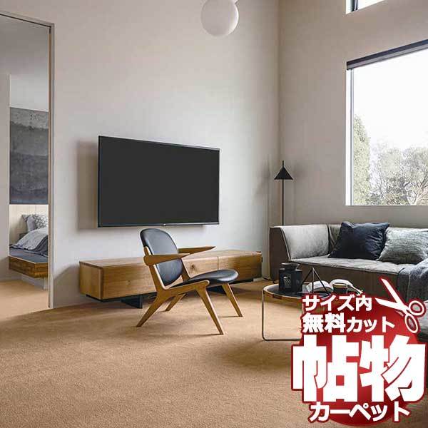
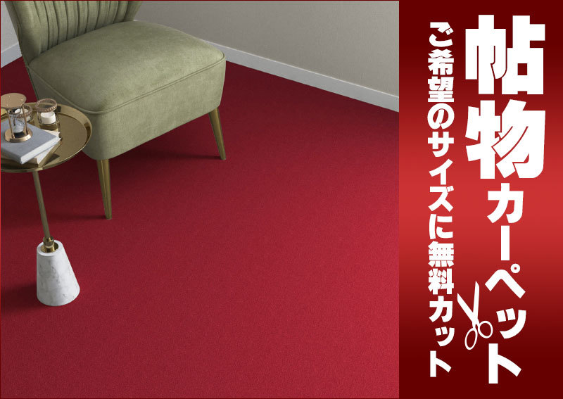
カーペット SG-6043 横240×縦240cm ロック加工
(税込) 送料込み
商品の説明
商品説明
オーダー カーペット 23270円カーペット SG-6043 横240×縦240cm ロック加工住まい、インテリア家具、インテリア
シンコール 日本製 カーペット
激安・サイズ内カット無料!
カーペットの踏み込む感触が明日を奏でる。
ホテルの客室や宴会場、各種施設から住宅まで幅広く使用できるカーペット。ロールカーペットの魅力とは、人と自然とが紡いできた「繊維の優しさ」そのものです。
シンコールでは、カーペットの繊維が織りなす優しさに包み込まれる心地よさ。シームレスに広がるデザイン。歩く度に、寝転ぶ度に感じるロールカーペットの魅力をお届けします。
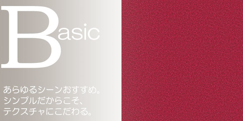
SG-6043(シグマ)
●組成:ナイロン100%
●防炎試験番号:E 2230242
●パイル長mm:カットパイル 7.0
●総厚mm:9.0
●ロール幅cm:364
●対応品番:SG-6039 SG-6040 SG-6041 SG-6042 SG-6043 SG-6044 SG-6045 SG-6046 SG-6047 SG-6048
シンコールロールカーペットで空間をデザイン
さまざまな人が集う空間を美しさと心地よさで満たす、シンコールロールカーペット。「清潔」「安全」「快適」「デザイン」といったロールカーペットには不可欠なエッセンスを妥協することなく追求した、圧倒的なクオリティをご紹介します。
オーダーカーペットとは
お客様のご希望に合わせて1cm単位で、お部屋にぴったりサイズでオーダーできるカーペットのことです。
場所を選ばないで、和風にも洋風にもピッタリのカーペットをお作りできます。 カーペットのカラーも豊富・サイズは、マット 玄関マット ラグ 廊下敷き 江戸間 本間 中京間 2畳 3畳 長4畳 4.5畳 長4.5畳 6畳 7.5畳 8畳 10畳 12畳 など豊富です。
丸型や変形カットも無料・カーペットの端部処理加工を選べるのはもちろん、防音性、抗菌・防臭・防汚加工などの機能性を重視したい方、オールシーズン使えるウール素材やお手入れのしやすいポリエステルなどあります。お客様のオリジナル好みに合わせてお届けする、それがオーダーカーペットです。
カーペットの敷き方はいろいろ
カーペットは防音効果がありますので、廊下への足音対策や冷え防止にもおすすめ。汚れに強いカーペットは食卓テープルの下に、抗菌・防ダニカーペットはお子様も安心してお使いいただけます。
※サイズ内カットの注意点
1.ご希望のカーペットを規格サイズの中から1cm単位でオーダーできます。
2.サイズ内オーダーカーペットは、直線カット1cm単位(但し切込みカットは5cm以上1cm単位)
3.カーペットは繊維製品です。1%程度の延び縮みが発生することもございますのでご了承ください。
4.お部屋には多少の変形がある場合もございます。オーダーカーペットは少し小さめの寸法でご注文いただくことをお奨めいたします。
5.カーペットの価格は(四角・丸形・変形とも)総巾・総高さのサイズに応じた価格になります。
6.ご指示いただきましたら、サイズ内カットした後、カーペットのカットロスを同送いたします。ただしカットロスの再カットはいたしません。また、ご指示のない場合は弊社にて処分させていただきます。
7.カーペットの周りを糸が解けないようきれいに加工するロック加工はご注文日より約3~5営業日でお届けいたします。


商品の情報
カテゴリー
配送料の負担
送料込み(出品者負担)配送の方法
ゆうゆうメルカリ便発送元の地域
宮城県発送までの日数
1~2日で発送メルカリ安心への取り組み
お金は事務局に支払われ、評価後に振り込まれます
出品者
スピード発送
この出品者は平均24時間以内に発送しています